Special Eye Testing
Optical Coherence Tomographer (OCT)
Our OCT helps us better manage glaucoma and diseases of the retina because this technology allows the eye doctor to see the deep tissue layers in the eye. Similar to ultrasound, this diagnostic technique employs light rather than sound waves to achieve higher resolution pictures of the structural layers of the back of the eye. These high-definition images are the only way that they can actually see beneath the surface to the nerve fiber layers where damage occurs. Up until now, eye doctors had to use other tests to indicate damage in this critical area of sight. Common eye diseases such macular degeneration, diabetic retinopathy, and glaucoma are detected early by the OCT when the diseases can be more effectively treated.
ZEISS / Humphrey Visual Field Testing
Visual Field testing can help save vision because it is another test used to diagnose or rule out glaucoma and other neurological disorders that affect vision. This simple, but effective service has saved lives by detecting various medical conditions such as strokes, brain tumors, and other neurological defects.
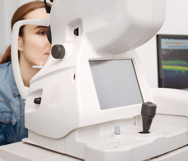

Zeiss Cirrus OTC
At 100,000 scans per second, ZEISS CIRRUS 6000 enables clinicians to image a larger field of view up to 12mm in a single scan. It also captures high-defintion (HD) OCT and OCT Angiography (OCTA) scans, revealing the finer microvascular details of the retina and providing more insight into your patient’s condition.
The CIRRUS platform offers clinicians extensive, clinically-validated applications—for retina, glaucoma and anterior segment—that allow for precise analysis, faster throughput, and smarter decision-making across a range of clinical conditions and patient types.
Essilor Visioffice Digital Measuring System
In Office Lens Finishing
Our special in-office lab can speed up your lens orders and provide lenses when you are in a pinch.
Today’s high-resolution lenses require laser-precision measurements to perform their best. Our Visioffice system from Essilor allows the opticians to obtain every possible parameter required for modern lenses with extreme accuracy. Our Visioffice system will let you see your best from your new custom, digital lenses from Varilux. No-line bifocals and single vision lenses can be produced precisely the way your eyes will be using them.
Our high-definition digital lenses use the Visioffice® System to calculate a perfect match of lenses based on eye, frame and behavior data. This “dynamic 4D” measurement system uses eye data by precisely tracking optical Eye Center Rotation, or “ERC.” To focus, the eye rotates around a fixed point called the ERC. The Visioffice® system uses advanced technology to measure this ERC and your dominant eye to make your custom lenses. Frame data such as the size and fit are taken into account to ensure that your new lenses are made precisely for your frame. Behavior data uses natural head posture and gaze to target best vision where you look and see. High-definition digital lenses uses the latest in manufacturing techniques to ensure sharper vision at any angle and under any lighting conditions.
Enhanced Fit™ lenses use W.A.V.E. technology™ to provide sharper vision as well as take into account frame data and prescription to make custom lenses for you. Ipseo™ lenses add behavioral data to create a lens that matches you personally and creates wide, comfortable vision for progressive lens wearers. Fit™ and Eyecode™ are available in multifocal and single vision versions. The result is clear, sharp vision all the time. Ask us how the Visioffice™ system can enhance your vision.
i.Care Tonometry
Tonometry is a test to measure the pressure of the fluid inside your eyes. It is an important test in the evaluation of patients at risk from glaucoma.
Our Icare tonometer is based on a proven accurate measuring principle, in which a very light probe is used to make momentary and gentle contact with the cornea. The measurement is barely noticed by the patient and often does not even cause corneal reflex.
Retinal Digital Imaging
A high-definition digital image of the retinal area helps your eye doctor diagnose and manage eye diseases in the delicate retinal area. Damage to these delicate structures of the retinal area is often the first sign of systemic diseases such as MS, diabetes and more. The retina is the “window to the body” and routine retinal imaging can help your eye doctor monitor the changes in your eye health from year to year.